
Presentation
Sudden onset of left sided body weakness.
Patient Data
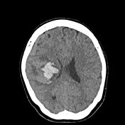

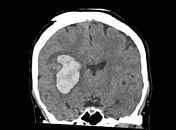
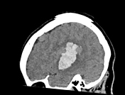

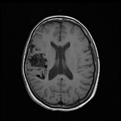
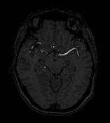
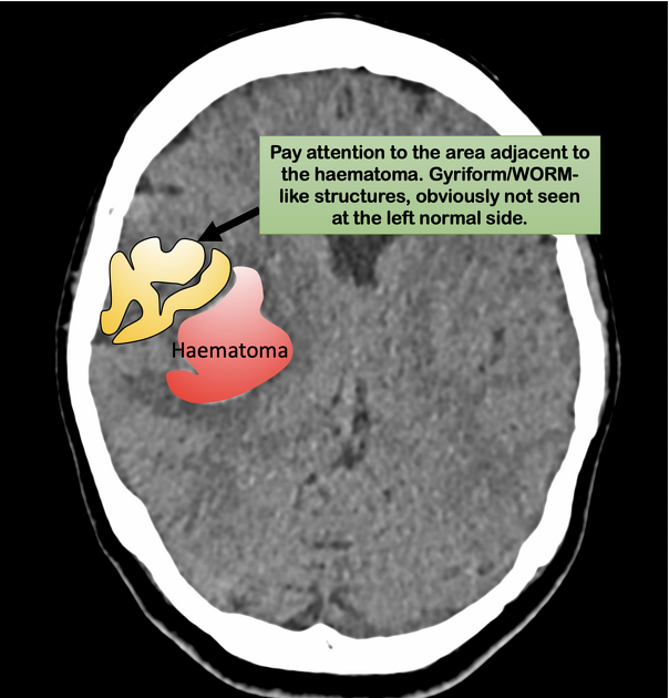
Acute intraparenchymal hematoma with epicenter at the right insula.
Abnormal gyriform-like/worm-like slightly hyperdense structures lateral to the aforementioned hematoma and within the right Sylvian fissure.
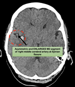
Asymmetrical dilatation of M1 and M2 segments of right middle cerebral artery as well as enlarged cortical veins at right subdural/subarachnoid spaces.
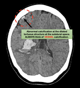
Abnormal calcification noted within the enlarged vessels at the right fronto-temporal region.
No significant midline shift or hydrocephalus.
Proceeded with MRI brain after 2 months of initial presentation.
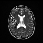
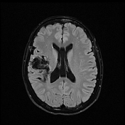

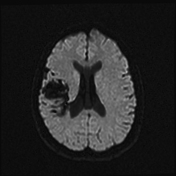
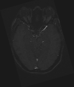
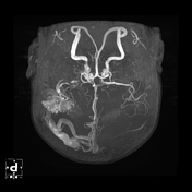
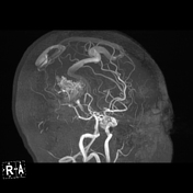

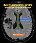
Serpiginous flow-voids at the right fronto-temporal region which represents the nidus of arteriovenous malformation.
Arterial supply is arising from the right middle cerebral artery, which the MCA itself is larger in caliber compared to left counterpart.
Multiple sites of draining veins, include dilated right vein of Trolard, right cortical veins draining into the superior sagittal sinus, another draining vein is via enlarged right basal vein of Rosenthal into the great vein of Galen.
No arterial aneurysm.

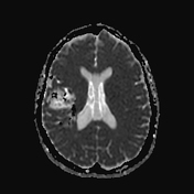
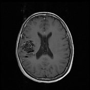
Annotated images showed some of the review areas for acute lobar hematoma in order to identify the possible diagnosis of arteriovenous malformation.
Case Discussion
Right fronto-temporal cerebral arteriovenous malformation. This case showed that the location of the acute intraparenchymal hematoma which is very near to the right basal ganglia/lentiform nucleus to be wrongly attributed to the TYPICAL location of hypertensive bleed. However, there are many imaging features to suggest the possibility of the cerebral AVM in plain CT brain.
- Abnormal vessel calcification adjacent to the acute hematoma
- Dilated and tortuous cortical veins
- Abnormal worm-like structure (not like normal cortex) adjacent to the acute hematoma.
Reporting radiologist should always look for the possible etiology for acute lobar hematoma.


 Unable to process the form. Check for errors and try again.