
Presentation
Cough for three days. Incidental finding.
Patient Data
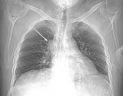


An enlargement of the azygos vein arch on the right side of the superior mediastinum (arrow) noted in the scanogram.
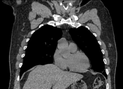
There is an interruption of the infrahepatic-suprarenal portion of the IVC associated with dilated hemiazygos vein, which drains into dilated azygos vein that ascends in its usual course in the posterior mediastinum until it joined the superior vena cava. The infrarenal IVC was not included in the examination.
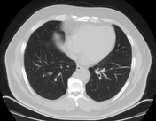
Atelectatic bands seen at the inferior lingula and left lower lung lobe.
A minute calcified nodule seen at the basal segment of the left lower lung lobe. Otherwise, normal both lungs.
Case Discussion
This case shows hemiazygos continuation of the IVC which was incidentally discovered during CT of the chest.
Although the infrarenal IVC was not included in the examination, we could expect its presence on the left side due to dilatation of the hemiazygos vein. If it was right-sided, it would drain directly into the azygos vein with no dilatation of the hemiazygos vein.
Also, double infrarenal IVC could be excluded due to the normal caliber of the azygos vein distal to the drainage of the hemiazygos vein.
So we finally could suggest the diagnosis of "left side IVC with hemiazygos continuation".


 Unable to process the form. Check for errors and try again.