
Presentation
Occasional dysphagia to solids
Patient Data


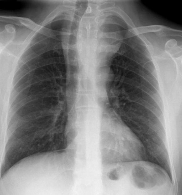
Chest x-rays demonstrate a large mass in the superior mediastinum, displacing to the right and somewhat narrowing the trachea. There is thickening of the right paratracheal stripe suggesting that it extends to the right of the trachea also. The outline of the mass fades out inferiorly, and does not obscure the arch or the aorta or the descending aorta.

The mass projects both to the left (orange) and right (yellow) the trachea (blue) which is displaced towards the right. The superior border of the mass fades out both superiorly and inferiorly but does not obliterated the outline of the aortic arch and descending aorta (green).
Case Discussion
This case illustrates the typical appearance of a large goiter extending behind the sternum. It was confirmed on CT and resected.


 Unable to process the form. Check for errors and try again.