
Presentation
Abdominal pain.
Patient Data
Age: 55 years
Gender: Male
From the case:
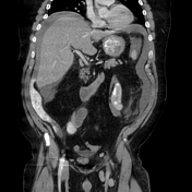
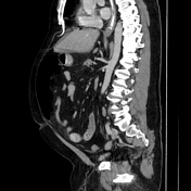
Perforated duodenal ulcer with leakage of oral contrast


Show annotations
Download
Info
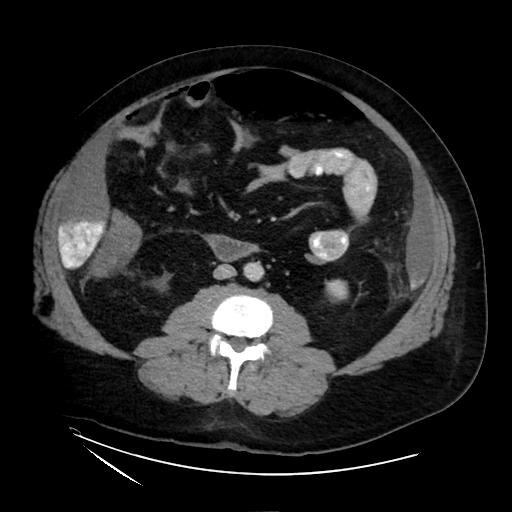
Nasogastric tube with oral contrast injected prior to the exam. Upper abdominal free air and fluid, with locules in the hepatic hilum/GB fossa. Dense oral contrast layering within the perihepatic fluid (along right lower margin and into the paracolic gutters R>L). Thickening of the first portion of the duodenum with a subtle wall defect indicating the ulcer channel.
Case Discussion
Perforated duodenal ulcer resulting in a large amount of leakage or oral contast into the peritoneal cavity. The defect is surprisingly subtle in this CT, however, which is fairly typical of ulcers due to the surrounding wall edema. This was confirmed at the time of surgery and repaired with Graham patch.


 Unable to process the form. Check for errors and try again.