
Presentation
Abdominal pain.
Patient Data
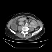

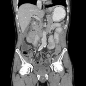
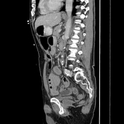


Free air and fluid in the upper abdomen anterior to the liver, heptic hilum and GB fossa. Subtle thickening of the duodenal bulb with a barely visible thin vertical channel.
Right lower quadrant peritonitis with irregularity and air adjacent to the appendix, suggesting perforation.
Case Discussion
This was a puzzling and unexpected case to report. The pattern of free air is very typical of a perforated duodenal ulcer, particularly when air locules and fluid are seen in the hepatic hilum and GB fossa. However, there was also RLQ peritonitis with and irregular appendix and extraluminal air. Thus perforated appendicitis was the favored diagnosis.
This patient went to the operating room where they surprisingly found a normal appendix with inflammatory debris/exudate in the RLQ, and a perforation of the duodenal bulb. Thus, it is suspected that the perforated ulcer resulting debris traveling to the RLQ, and this serves as a good warning that the typical pattern of free air from ulcers should not be ignored! It is challenging to conceive of not favoring appendiceal perforation given the amount of air/debris next to the appendix, but this is a good reminder that appendiceal and sigmoid diverticular perforations usually result in localized collections and air in the lower abdomen that dissect into the mesentery, and only rarely cause free air in the upper abdomen like seen in this case.


 Unable to process the form. Check for errors and try again.