
Presentation
Palpable lumps at the left lumbar region and right iliac fossa. History of myomectomy.
Patient Data


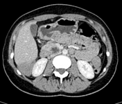
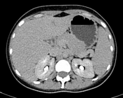
Multiple peritoneal solid masses are seen as follows:
-In the hepatic fissure for ligamentum teres.
-Multiple amalgamated masses along the para colic gutters at both sides. The one on the right side is extending upwards into the Morison pouch with scalloping of the posterior surface of the liver.
-Other discrete lesions in the pelvis.
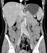
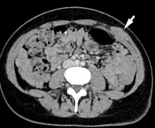
Similar lesion is seen in the anterior abdominal wall at the left lumbar region between the external oblique and internal oblique muscles.
All lesions show homogenous attenuation with increased enhancement in the delayed phase.
Minimal ascites is noted
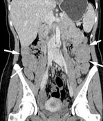
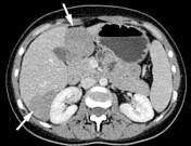

The first two images show the peritoneal masses.
The third image shows the abdominal wall mass.
Pathology report
Clinical data:
Biopsy from abdominal wall mass and peritoneal nodules with history of leiomyoma.
Gross examination:
Received multiple cres, all processed.
Microscopic examination:
Examination of samples from all sites revealed multiple interlacing bundles of bland non-dividing smooth muscle fibers with intervening fibrovascular stroma. No mitosis, no necrosis, no malignancy.
Diagnosis:
Picture is consistent with leiomyomatosis peritonealis disseminata.
Case Discussion
Multiple peritoneal masses in a young woman, pathologically proven to be diffuse peritoneal leiomyomatosis, a.k.a. leiomyomatosis peritonealis disseminata, most probably associated with the prior myomectomy.


 Unable to process the form. Check for errors and try again.