
Presentation
Increasing head circumference.
Patient Data


MRI of the brain of a child demonstrates an abnormal posterior fossa. The vermis is hypoplastic and rotated upwards, below which a large cystic space continuous with the fourth ventricle is present. The posterior fossa is elevated. The aqueduct appear narrowed and there is prominent obstructive hydrocephalus. The over-all size of the skull is increased.

A flow void is now seen through the floor of the third ventricle with reduced dilatation of the third and lateral ventricles (implied by reduction in upward bowing of the corpus callosum).

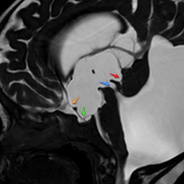
Pre third ventriculostomy there is marked distention of the third ventricle with ballooning out of the recesses:
- supraoptic (orange arrow)
- infundibular (green arrow)
- pineal (red arrow)
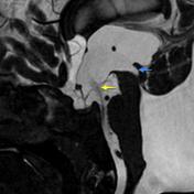
Post third ventriculostomy has been performed with flow seen through the floor of the third ventricle (yellow arrow). Funnelling of the superior part of the aqueduct (blue arrow) is seen in keeping with aqueduct stenosis due to extrinsic compression.
Case Discussion
This case illustrates a classic Dandy Walker malformation with associated aqueduct stenosis and obstructive hydrocephalus, a common associated feature (although not part of the triad 1. hypoplasia of the vermis with cephalad rotation, 2. cystic dilatation of the fourth ventricle, 3. enlarged posterior fossa )


 Unable to process the form. Check for errors and try again.