
Presentation
Decreased visual acuity for 1 month.
Patient Data


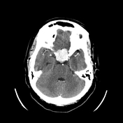
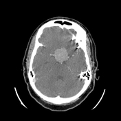
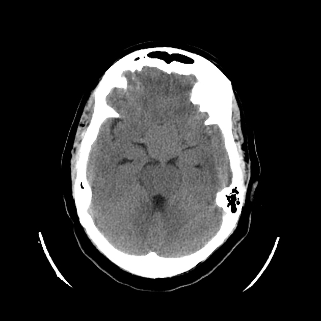
Suprasellar mass compression the optic chiasm.
Isoattenuating to brain parenchyma on uncontrasted scan.
Vivid enhancement.
Type of specimen
Suprasellar mass
Gross examination
The specimen consists of multiple fragments of brown tissue, blood and gauze with a combined weight of 9.6 g. The largest fragment measures 26 x 12 x 10 mm. All tissue processed.
Microscopic examination
Microscopic examination shows abundant tumor with a microcystic appearance. Fragments of blood and cauterised tissue are also noted. The tumor cells are predominantly elongated with vacuolated cytoplasm, resembling microcysts. Admixed nests of epithelioid cells arranged in meningothelial whorls are interspersed between the microcystic areas. These cells are arranged in a syncytial fashion, with indistinct cell membranes and eosinophilic cytoplasm. The nuclei are round and uniform with focal nuclear pseudoinclusions. No psammoma bodies are identified.
In the presence of adequate positive and negative controls, immunohistochemical stains confirm the presence of a meningioma (ER positive in 90% of tumor cells, EMA diffusely positive, D2-40 focally positive, S-100 protein negative and GFAP negative). Ki-67 is positive in <1% of tumor cells, indicating a low proliferative index. PAS is negative and alcian blue highlights acid mucin within the microcystic spaces.
Morphological, histochemical and immunohistochemical features are consistent with a microcystic meningioma.
Diagnosis Suprasellar mass: Microcystic meningioma (WHO grade 1)
Case Discussion
Suprasellar mass causing optic chiasm compression and decreasd visual acuity. The differential included pituitary macroadenoma and meningioma. Vivid enhancement suggested meningioma, confirmed on histology.


 Unable to process the form. Check for errors and try again.