
Presentation
Seizures.
Patient Data
Age: 35 years
Gender: Male
From the case:
Arteriovenous malformation - cerebral


Download
Info
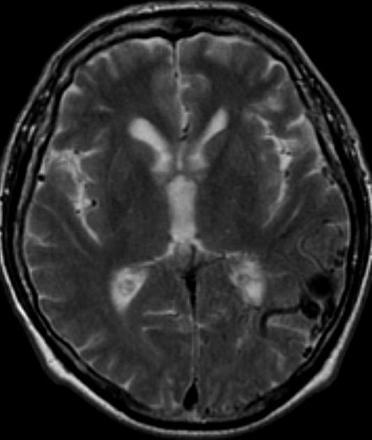
Axial T2 image shows multiple serpiginous vascular channels are noted involving the left temporoparietal brain parenchyma.
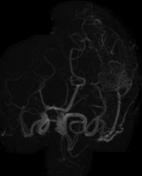
MRA and MRV images show multiple arterial feeders from the M4 branches of the left MCA and multiple dilated draining veins into superficial cortical veins consistent with arteriovenous malformation (AVM).
Case Discussion
This case represents a classic form of arteriovenous malformation (AVM). The flow voids are easily seen on T2 images. MR angiography shows the feeding arteries and draining veins, yet digital subtraction angiography remains the gold standard for AVM assessment; detecting the location and number of feeding vessels and the pattern of drainage.


 Unable to process the form. Check for errors and try again.