
Presentation
Chest pain and cough.
Patient Data
Age: 40 years
Gender: Male
From the case:
Pleural lipoma
Download
Info
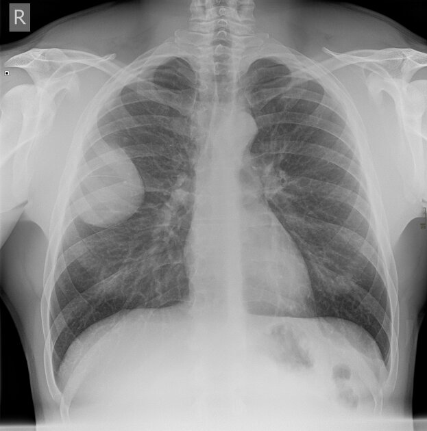
8 cm right upper thoracic chest wall mass.
Heart size normal. Lungs clear. Normal mediastinum.
From the case:
Pleural lipoma

Download
Info
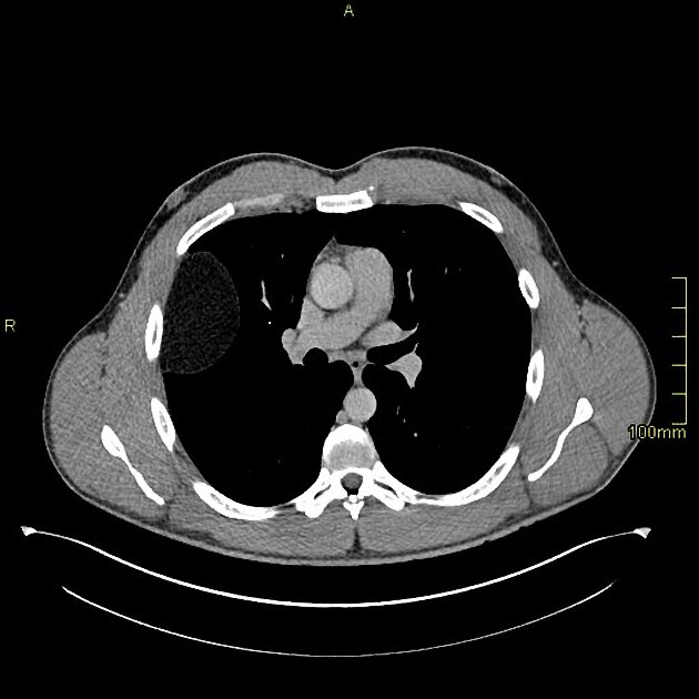
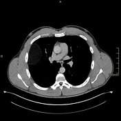
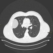
8 cm right fatty pleural based mass. No calcification.
Normal mediastinum. Lungs clear.
Case Discussion
Not all masses on chest radiographs are cancer.
The chest x-ray is a good example of identifying if a mass is pleural or lung based with the obtuse angle between the mass and chest wall indicating a non-lung origin.


 Unable to process the form. Check for errors and try again.