
Presentation
Painless testicular swelling on right side for one month. Some discomfort, otherwise no symptoms.
Patient Data

A large heterogeneous solid-cystic mass lesion is seen in the right scrotal sac, and right testis could not be seen separately from the lesion. It shows internal vascularity.


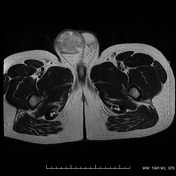
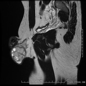
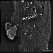

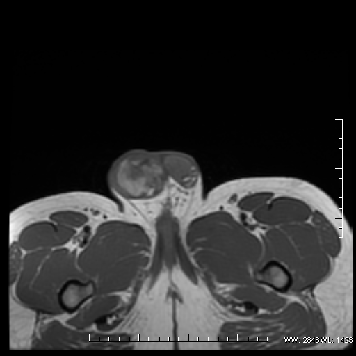
Well defined heterogeneous solid mass lesion is seen in right testis, with solid and cystic areas within, with possible hemorrhagic areas within.
Immediately after MRI, CT was done as correlative imaging and specifically to rule out calcification (teratoma/carcinoma).
Solid-cystic mass lesion in right testes. MRI gadolinium contrast is still seen. No calcification is however seen.
Note: This case has been tagged as "legacy" as it no longer meets image preparation and/or other case publication guidelines.
Case Discussion
Characteristic heterogeneous appearance, with cystic areas, in testicular mass lesion, leads to a provisional diagnosis of non-seminomatous germ cell tumor. On further investigation, his serum AFP and B-HCG were found to be elevated. Patient underwent orchiectomy with a pathological diagnosis of non-seminomatous mixed germ cell tumor


 Unable to process the form. Check for errors and try again.