
Presentation
Bitemporal Hemianopsia.
Patient Data
Note: This case has been tagged as "legacy" as it no longer meets image preparation and/or other case publication guidelines.

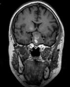
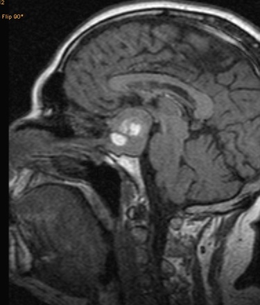
Large sellar and suprasellar mass lesion with homogenous texture showing multiple cystic changes with high T1W signals reflecting intratumoral hemorrhage. The pituitary gland is not identified with widening of the sellar floor which remains intact in keeping with pituitary origin.
It is extending to the suprasellar cistern elevating and compressing the optic chiasm. Also, laterally displacing both cavernous sinuses, with partial encasement of both internal carotid arteries.
Case Discussion
This case proved to be pituitary macroadenoma with intratumoral hemorrhage, also known as pituitary apoplexy.


 Unable to process the form. Check for errors and try again.