
Presentation
Abdominal pain and distention with nausea.
Patient Data
Age: 50 years
Gender: Female
From the case:
Cecal volvulus

Download
Info
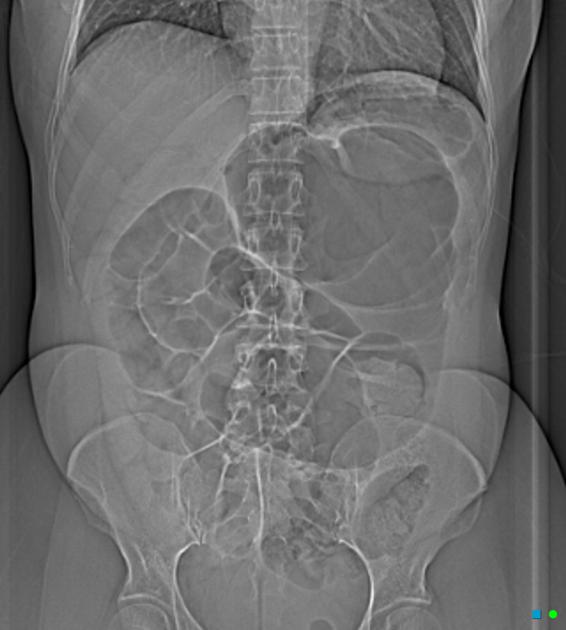
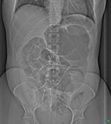
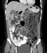
Scout view showing a markedly distended large bowel loop in the left side of the abdomen. CT demonstrates the large bowel mass is a grossly distended food-filled cecum that is not in the right iliac fossa but in the left side of the abdomen. The axial slices clearly show the ileum passing through a 360-degree rotation to enter the abnormally positioned cecum.
From the case:
Cecal volvulus
Download
Info
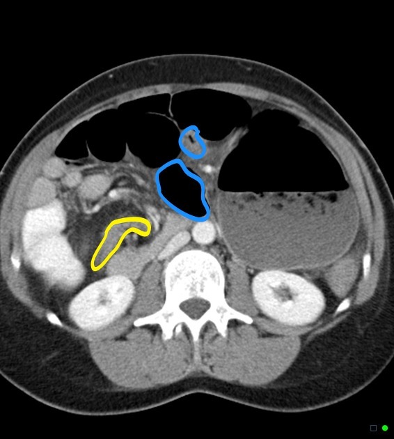
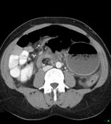
The terminal ileum (yellow) and ascending colon (blue) can be traced to whirl around each other, the mesenteric vessels (red) acting as the axis of rotation. The appendix is also visible, distended by gas and fluid.
Case Discussion
This case illustrates fairly typical appearances of a cecal volvulus.


 Unable to process the form. Check for errors and try again.