
Presentation
Incidental findings during workup for abdominal pain. Deranged urea and electrolytes.
Patient Data


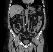
Multiple incidental findings:
duplicated IVC
cystitis cystica
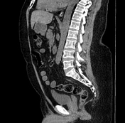
type 1 fixed hiatal hernia
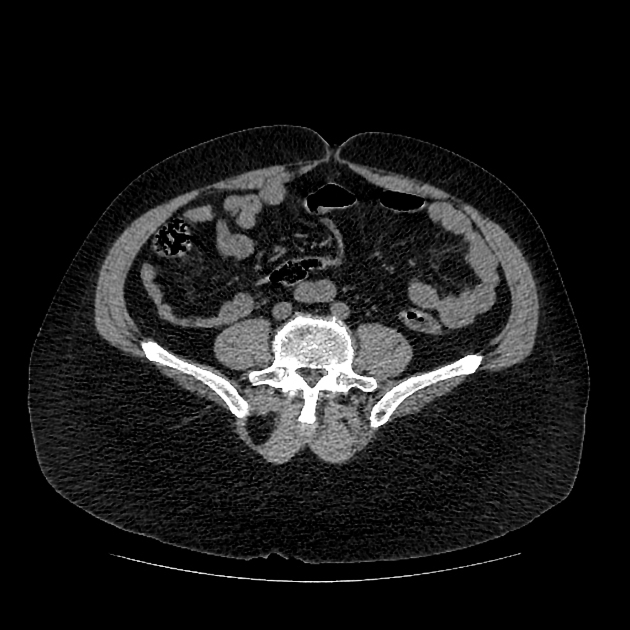
right erector spinae intramuscular lipoma
Case Discussion
There is a duplicated IVC as demonstrated by the continuation of the tortuous left common iliac vein and crossing anterior to the aorta at the level of the left renal vein to join the normal right-sided IVC. There are no associated renal parenchymal or vascular anomalies in the non-contrast CT study.
There is the appearance of multiple, tiny, lucent bladder wall defects suggestive of cystitis cystica. There is no associated prostatomegaly, no bladder calculi, and no pelvic lipomatosis. Chronic infection and irritation as the possible source was suggested.
Incidental, simple, paraspinal intramuscular lipoma for conservative management.


 Unable to process the form. Check for errors and try again.