Patient Data
Note: This case has been tagged as "legacy" as it no longer meets image preparation and/or other case publication guidelines.


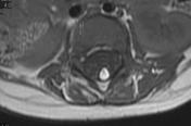
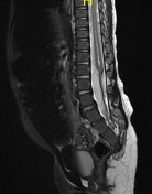
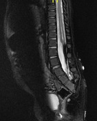
Partial agenesis of sacrum seen. Note the filum terminale lipoma which is suppressed on this fat sat image. Note the "club" shaped conus with syrinx - the club-shaped blunted conus is typical for sacral agenesis.
Case Discussion
There are four types of sacral agenesis:
In type 1, partial unilateral agenesis is localized to the sacrum or coccyx.
In type 2, there are partial but bilaterally symmetric defects in the sacrum. The iliac bones articulate with S1, and distal segments of the sacrum and coccyx fail to develop.
In type 3, there is total sacral agenesis and the iliac bones articulate with the lowest available segment of the lumbar spine.
In type 4, there is total sacral agenesis and the iliac bones are fused posteriorly along the midline.


 Unable to process the form. Check for errors and try again.