
Presentation
Chronic perianal infection
Patient Data
Age: 45 years
Gender: Female
Note: This case has been tagged as "legacy" as it no longer meets image preparation and/or other case publication guidelines.
From the case:
Perianal fistula - grade I


Download
Info
Coronal contrast-enhanced MR image shows the fistula with contrast enhancement
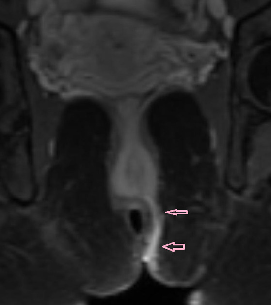
Axial contrast-enhanced MR image shows the fistula and inflammatory tissue within the left intersphincteric plane.
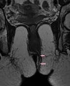
Coronal T2SE MR image shows a fistula (arrow) within the left inter-sphincteric plane. Note the entry site in midline in the lower third of the anal canal.
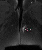
Axial T2SE MR image shows the fistula (arrow) as a dot.
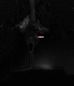
Axial T2SE MR image higher shows the fistula approaching the midline at 12 hour which is unusual.
Case Discussion
MRI often does not shows clearly the entry point of the fistula.


 Unable to process the form. Check for errors and try again.