
Presentation
Patient complaints heaviness of the right upper abdomen since long time
Patient Data


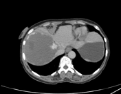

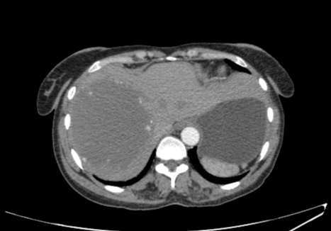
The liver is normally positioned and has normal size. There is a large, definable, mass lesion of about 9.7 x 9.3 x 11.8 cm (AP x TRANS x CC) in the right lobe of the liver parenchyma which is showing nodular and non-continuous, peripheral enhancement on arterial phase and progressive peripheral enhancement with more centripetal fill-in on venous and delayed phases suggestive of giant hepatic hemangioma.
At least two small, well-defined, hypodense lesions are also seen in the left lobe of the liver which are most likely simple cysts (as they show no definite density changes on provided phases). The hepatic and portal veins are patent. The intra hepatic and extra hepatic bile ducts are unremarkable.
Multiple dominant lymph nodes are noted in pre/para-aortic and aorto-caval regions which are most likely due to infective/inflammatory disease process.
Images through chest were with in normal limits (not shown).
Case Discussion
Patient has done abdominal USG by which it was labeled as hepatic mass lesion and further work up was advised, so after doing CT Tri-phasic abdomen and pelvis findings were in favor of giant hepatic hemangioma with most likely hepatic cysts and abdominal lymphadenopathy as described above.


 Unable to process the form. Check for errors and try again.