
Presentation
Long standing swelling medial to left eye. CT scan outside reported as "orbital dermoid".
Patient Data
Age: 10 months
Gender: Male
Note: This case has been tagged as "legacy" as it no longer meets image preparation and/or other case publication guidelines.
From the case:
Nasal encephalocele

Download
Info

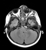
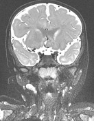
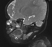
There is a cystic lesion medial to the left globe. Coronal images demonstrate continuity of the cyst with a herniated frontal lobe tissue and the intracranial compartment.
Case Discussion
Encephaloceles are uncommon, but are important lesions to consider. Imagine the results of deciding to excise this 'dermoid' without appreciating the actual pathology.


 Unable to process the form. Check for errors and try again.