Presentation
Known avascular lesion in the sella turcica followed with MRI for more than 4 years.
Patient Data


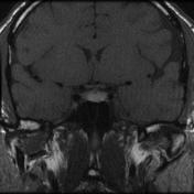

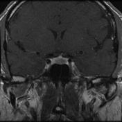

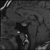

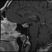

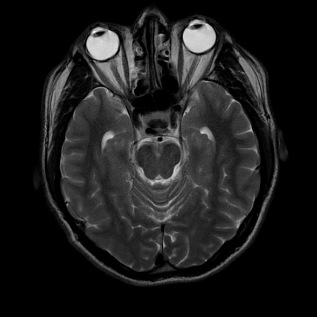
Interestingly, besides the T2 hypo and T1 hyperintense Rathke cyst, which does not enhance, there is also a Thornwaldt cyst in the epipharynx. Both Rathke's pouch and Thornwaldt cysts (epipharyngeal bursae) are closely related to the chorda dorsalis during embryogenesis.
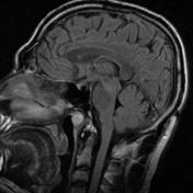
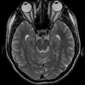
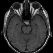
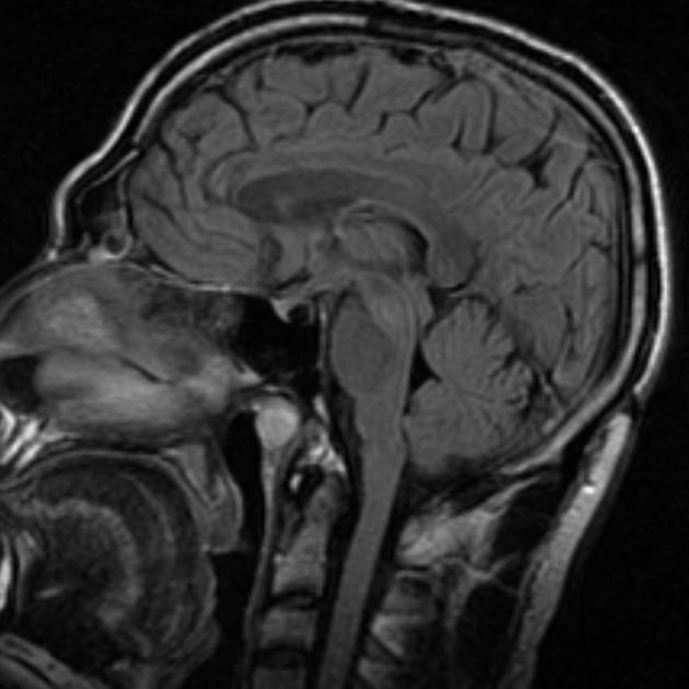
T2 hypointense and T1 hyperintense lesion in the sella, anterior to the neurohypophysis, which also shows T1-hyperintense (but not T2 hypointense) signal.
Case Discussion
T2 hypointensity in a pituitary cyst is regarded as highly suggestive of a Rathke cleft cyst 1.


 Unable to process the form. Check for errors and try again.