
Brain metastases from lung cancer associated with metastasis to the pineal gland
Presentation
Known lung cancer. Behavioral changes with confusion.
Patient Data


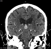
Multiple intra-axial cerebral masses, both supratentorial and infratentorial, exhibit solid enhancement, with some displaying a ring-enhancing pattern associated with central necrosis.
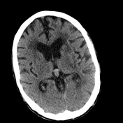
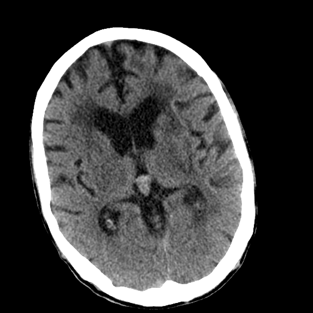
The pineal mass appears spontaneously mildly hyperdense and enhanced following contrast media administration.
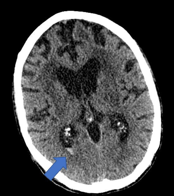
A thin layer of intraventricular hemorrhage is observed in the occipital horn of the right lateral ventricle.

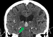
The pineal mass appears enhanced following contrast media administration (green arrow).
A thin layer of intraventricular hemorrhage (blue arrow) in the occipital horn of the right lateral ventricle.
Case Discussion
Pineal gland metastasis is an uncommon site for metastatic disease.
In our case, it is challenging to determine the extent of a mass effect on the ventricular system, given the patient's advanced age, which is accompanied by cerebral atrophy and ventricular ectasia, along with periventricular hypodensities. This situation leads us to consider two hypotheses: either ex vacuo hydrocephalus associated with white matter lesions or obstructive hydrocephalus with minimal transependymal resorption.
The absence of effacement of adjacent cerebral sulci favors an ex vacuo hydrocephalus.


 Unable to process the form. Check for errors and try again.