
Presentation
Complained of weakness in the left half of her body. Routine blood workup revealed hyponatremia and was treated with correction. After 10 days, in view of the history of left-sided weakness which partially improved, she underwent an acute stroke CT/MR protocol.
Patient Data
Note: This case has been tagged as "legacy" as it no longer meets image preparation and/or other case publication guidelines.
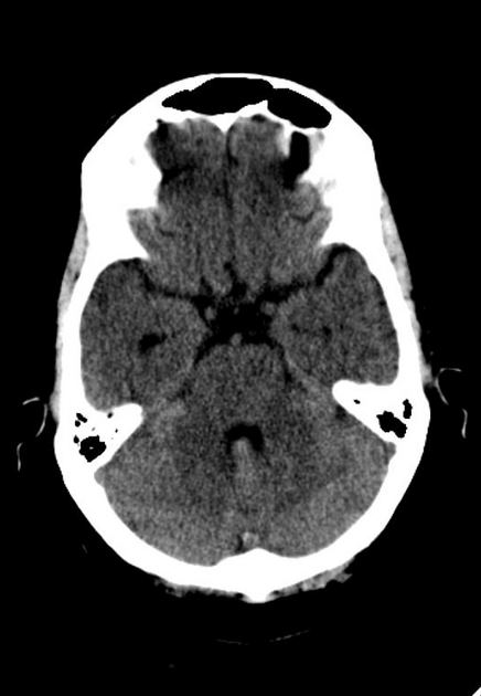
Diffuse hypodense areas seen involving bilateral deep nuclei and pons.


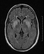
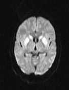
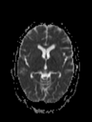
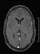
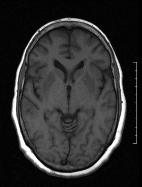
T2 bright signals from bilateral caudate head and basal ganglia, central pons with diffusion restriction. The sparing of ventrolateral pons is characteristic.
Case Discussion
On viewing the imaging findings, history of recent treatment was looked into. Hyponatremia at time of admission was 104 mEq, corrected to 121 mEq within 24hrs.
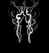
In the background of such a history with normal MRA, a diagnosis of central pontine myelinolysis with extra pontine myelinolysis involving bilateral basal ganglia was made. These conditions are now grouped under the term osmotic demyelination syndrome.


 Unable to process the form. Check for errors and try again.