
Presentation
Patient with sinus node disease, atrial fibrillation and hypertension. Scheduled for placement of a cardiac pacemaker. CT of the chest was requested to investigate possible interstitial associated pneumonia.
Patient Data
Age: 80 years
Gender: Female

Download
Info

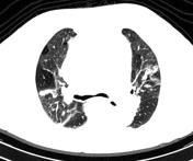
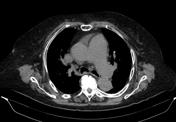
The lungs show a pattern of mosaic attenuation. Cardiomegaly and dilated pulmonary arterial vasculature.
Case Discussion
This case illustrates mosaic attenuation pattern presumed due to the known chronic pulmonary hypertension, which is secondary to heart disease.


 Unable to process the form. Check for errors and try again.