
Presentation
Abdominal pain and distension.
Patient Data

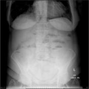
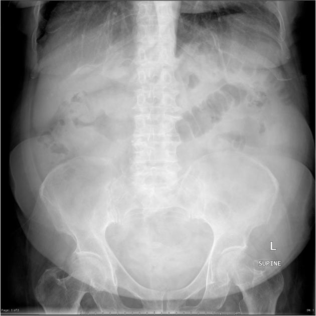
Multiple air fluid levels are present. A soft tissue mass appear present in the right upper quadrant with central air attenuation.


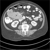
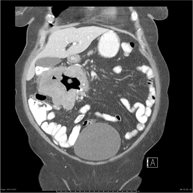
A large lobulated irregularsoft tissue mass is situated in the right half of the abdomen. It appears toarise from the junction of second and third parts of the duodenum and tocontain a cavity partially filled with contrast material and air. Itmeasures 11.7 x 6.9 x 9 cm which is significantly larger than at the time ofthe previous study where it was reported as measuring 3 x 4 x 4 centimeters.
No focal lesions are seen in the liver, spleen, pancreas or adrenals. A 6cm cyst arises from the mid pole of the right kidney. The kidneys areotherwise normal in appearance. There is no retroperitoneal or pelvic lymphnode enlargement & no free fluid. Incidental findings of a small paraumbilical hernia and a small hiatus hernia are noted. No focal bonylesions.
Patient went on to have a biopsy.
MICROSCOPIC DESCRIPTION
The sections show fragments of a tumor comprising spindle cells with plump, frequently cigar-shaped, nuclei, arranged in sweeping fascicles. There is moderate nuclear pleomorphism but only 1 mitosis per 50 high power fields. There are areas of hemorrhage within the tissue fragments (which may represent surgical artifact) and one fragment shows a single small focus of necrosis. The lesion contains a light, diffuse infiltrate of lymphocytes and plasma cells and in some areas there are a few neutrophils. Immunoperoxidase stains show that the tumor cells are positive for smooth muscle actin and negative for S-100 and c-Kit +ve.
FINAL DIAGNOSIS
Gastrointestinal stromal tumor of uncertain malignant potential showing smooth muscle differentiation.


 Unable to process the form. Check for errors and try again.