
Cephalisation of pulmonary veins in congestive cardiac failure
Diagnosis almost certain

Download
Info

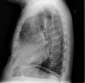
Frontal and lateral chest radiograph demonstrates cephalisation (upper lobe vascular redistribution) in stage I CCF where there is elevation of the left atrial pressure 10-15 mmHg. Normal left atrial pressure is 5-10 mmHg.
Download
Info

Determining cephalisation must be taken with caution. Comparison between the upper and lower lobe vessels is made at equal distance from the hilar point. The hilar point/angle is formed by the superior pulmonary vein (yellow line) and the descending pulmonary artery (green line).


 Unable to process the form. Check for errors and try again.