
Presentation
Patient with known neurofibromatosis type 1. She presented with cafe-au-lait spots shortly after birth. The patient was otherwise healthy.
Patient Data
Note: This case has been tagged as "legacy" as it no longer meets image preparation and/or other case publication guidelines.

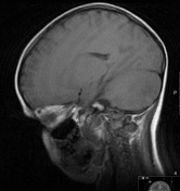
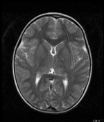

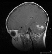
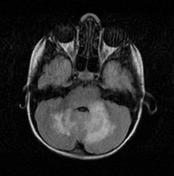

T2 MRI shows hyperintense lesions in the cerebellum. Some minor foci are also present in the basal ganglia. The lesions are isointense on T1-weighted images and hyperintense on T2-weighted and FLAIR images. The lesions do not show mass effect but enhance after contrast administration.
Case Discussion
The previous year a brain MRI showed focal areas of signal intensity (FASI) in the cerebellum. Follow-up CT showed contrast enhancement of the cerebellum lesions.
Astrocytomas are seen in 1-3% of NF 1 patients. Common sites are brainstem/cerebellum and splenium of corpus callosum. These tumors have an earlier presentation, are more likely multicentric and are much less aggressive than their sporadic counterparts. MR Spectroscopy may show increased choline in both FASI and tumors, but NAA is near normal in FASI while tumors show decreased levels. Persistent enhancement and increasing size are worrisome features for tumors.


 Unable to process the form. Check for errors and try again.