
Presentation
Infant with a right epicanthal vascular lesion.
Patient Data
Age: infant
Gender: Male
From the case:
Infantile hemangioma (periorbital)


Download
Info
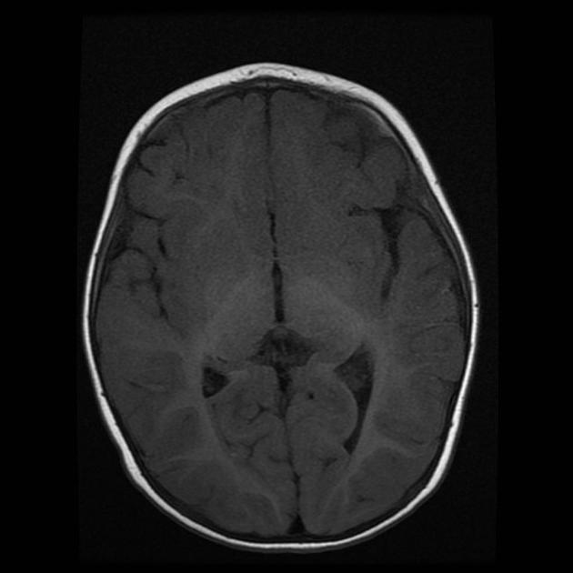
The findings are compatible with capillary hemangioma of the orbit. This one is smaller, located near the right epicanthal fold, with no intra-orbital extension. Hypointense on T1 and hyperintense on T2 with small flow-voids.
Case Discussion
Differential diagnosis for this lesion includes:
- arteriovenous malformation & lymphatic malformations
- meningocele
- very rarely - rhabdomyosarcoma or angiosarcoma
Related article:


 Unable to process the form. Check for errors and try again.