
Presentation
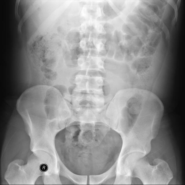
Right flank pain to rule out renal stones.
Patient Data
Age: 25 years
Gender: Male

Download
Info
An inflamed appendix with appendicolith is seen subjacent to the right hepatic lobe.
Download
Info
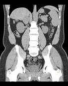
6.7 x 5.2 cm hyperechogenic lesion posterior to the right hepatic lobe with echogenic rim.
Download
Info

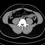
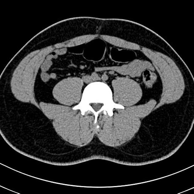
A subhepatic collection containing gas and appendicolith, in keeping with an appendicular abscess.
Case Discussion
Appendicular abscess is considered the most common complication of acute appendicitis, in particular after a perforated appendix.


 Unable to process the form. Check for errors and try again.