
Presentation
Mixed conductive/sensorineural hearing loss, since early childhood.
Patient Data


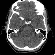

Dilated vestibular aqueducts.
Both cochlea are slightly globular (left worse than right) raising the possibility of cochlear dysplasia (modiolus present though).
The horizontal semicircular canal is slightly dilated bilaterally. The horizontal semicircular canal is the last canal to form and often the first to go, in terms of development.
Incidental likely calcified aneurysms - based on this finding, an MRI was performed
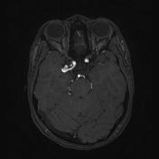

TOF MRA shows multiple aneurysms; right M1, right MCA bifurcation, right ICA paraclinoid, and left PCOM. There is no known association between LVAS and berry aneurysms.
A tortuous clump of vessels affecting the right PCA mimics and aneurysm.
Vessels are tortuous.
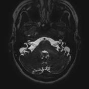
Large vestibular aqueduct syndrome; note how the endolymphatic sac lies outside the dura on the steady-state sequences.
Case Discussion
This case shows bilateral large vestibular aqueduct syndrome, multiple intracranial aneurysms and dolicoectasia. Tortuous vessels are seen in connective tissue diseases like Erlers Danhos, and fibromuscular dysplasia. There is no known association between LVAS and a connective tissue vasculopathy.


 Unable to process the form. Check for errors and try again.