
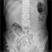
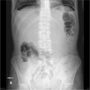

Free intraperitoneal gas is demonstrated. Bowel loops remain within normal limits. No destructive bony lesion.


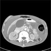
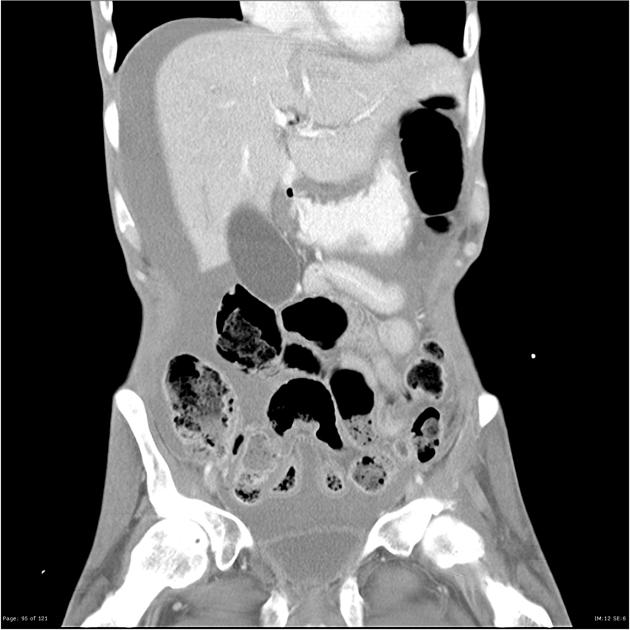
There is extensive free intraperitoneal fluid and gas. There is contrast extravasation along the greater curvature of the stomach in the region of the body/pylorus of the stomach, with contrast seen pooling in the fossa of the ductus venosum of the liver. Distal loops of small bowel and large bowel are nondilated. The liver, gallbladder, kidneys, adrenal glands, pancreas and bladder are within normal limits.
There is abnormal contrast 'swirling' within CFV, EIV and IVC most likely artefactual. However, an ultrasound of the thigh is suggested to exclude a DVT.
No destructive bony lesion. Lung bases are clear.
The patient went on to have a laparotomy, biopsy and closure of the perforated gastric ulcer.
Histology
MACROSCOPIC DESCRIPTION: "Four quadrants of gastric ulcer": Four biopsies 2-4mm.
MICROSCOPIC DESCRIPTION: Multiple levels examined show four small gastric biopsies. There is ulceration of gastric mucosa with near complete destruction of normal architecture. The ulcer base is covered by amorphous pink debris and the underlying mucosa contains apoptotic cells and granulation tissue composed mainly of lymphocytes and small regenerating vessels. There are few scattered normal glandular structures within the mucosa which lack atypia. Helicobacter are not seen. There is no evidence of dysplasia or malignancy.
FINAL DIAGNOSIS: Ulcerated and inflamed gastric mucosa
Case Discussion
Perforated gastric are relatively uncommon now that powerful acid suppression and Helicobacter pylori eradication is available.


 Unable to process the form. Check for errors and try again.