Presentation
Swelling at nape of neck region.
Patient Data
![](https://prod-images-static.radiopaedia.org/images/2635814/79fabebadab0732397f34b0ae3037d_thumb.jpg)
![This study is a stack](/packs/stack-YQKLCKBI.gif)
![](https://prod-images-static.radiopaedia.org/images/2635853/9152187f6638877c9db78c611e611a_thumb.jpg)
![This study is a stack](/packs/stack-YQKLCKBI.gif)
![](https://prod-images-static.radiopaedia.org/images/2635873/9d52cd83c88f707de1cbbdc2de9dc8_thumb.jpg)
![This study is a stack](/packs/stack-YQKLCKBI.gif)
![](https://prod-images-static.radiopaedia.org/images/2636014/61e1377a0a903c04b512e157b5b7bf_thumb.jpg)
![This study is a stack](/packs/stack-YQKLCKBI.gif)
![](https://prod-images-static.radiopaedia.org/images/2636004/dc93c7e952e890619e80bff7398330_thumb.jpg)
![This study is a stack](/packs/stack-YQKLCKBI.gif)
![](https://prod-images-static.radiopaedia.org/images/2636020/bc21af49e921b09551c6bb7f18cd2a_thumb.jpg)
![This study is a stack](/packs/stack-YQKLCKBI.gif)
![](https://prod-images-static.radiopaedia.org/images/2636093/86b15c4e76e54447f066a1562fc5cd_thumb.jpg)
![This study is a stack](/packs/stack-YQKLCKBI.gif)
![](https://prod-images-static.radiopaedia.org/images/2636143/9e71598c91b49a438c2c185496bd85_thumb.jpg)
![This study is a stack](/packs/stack-YQKLCKBI.gif)
![](https://prod-images-static.radiopaedia.org/images/2635814/79fabebadab0732397f34b0ae3037d_big_gallery.jpg)
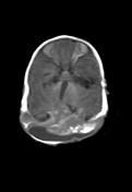
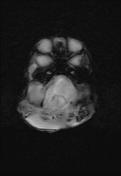
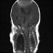
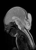
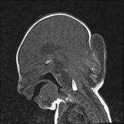
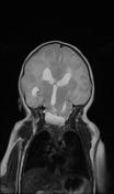
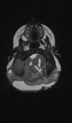
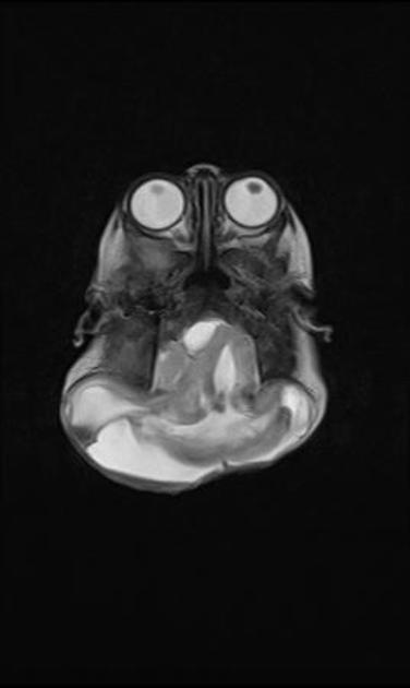
There is approximately 3.2 cm sized body defect seen at occipital bone with approx 6.3 (SI) x 2.9 (AP) x 7.8 (TR) cm sized herniated sac noted. There is herniation of occipital lobe, occipital horn of lateral ventricles, fourth ventricle, part of corpus callosum, both cerebellum and surrounding meninges. These represent occipital meningo-encephalocele.
The cerebellar hemisphere is probably disorganized. Multiple patchy areas of T1W hyperintensity seen within the herniated brain parenchyma, which shows blooming on GRE images, represent hemorrhage. Linear T1W hyperintensity seen anterior to the medulla measuring 2.0 (TR) x 1.0 (AP) x 2.1 (SI) cm in size, represents hemorrhage. There is widening of upper cervical canal.
There is hyperintense signal seen on the T2W and FLAIR images in the occipital lobes, representing gliosis.
The confluence of venous sinuses at torcula is not distinctly visualized.