
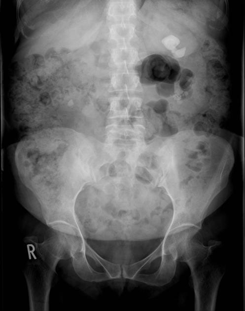
Presentation
Recurrent renal stone disease
Patient Data
Age: 45 years
Gender: Female
From the case:
Parathyroid adenoma - ectopic
Download
Info
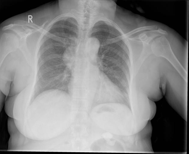
A chest radiograph was performed prior to percutaneous nephrolithotomy (PCNL).
Right sided mediastinal mass, which demonstrates the hilum overlay sign.
From the case:
Parathyroid adenoma - ectopic
Download
Info
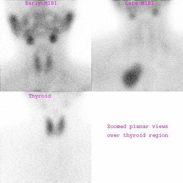
A MIBI scan was undertaken to assess for a parathyroid adenoma.
Avid tracer uptake in the mediastinal mass visualized on plain chest radiograph, confirms it represents an ectopic parathyroid adenoma.
Case Discussion
In a small number of cases, recurrent renal stone diseases (nephrolithiasis) is due to an underlying metabolic condition. This includes primary hyperparathyroidism due to a parathyroid adenoma. Rarely these occur in an ectopic location, typically the mediastinum, rather than in the neck.


 Unable to process the form. Check for errors and try again.