
Presentation
Incidental finding
Patient Data
Age: 40 years
Gender: Female
From the case:
Duplication of the inferior vena cava


Download
Info
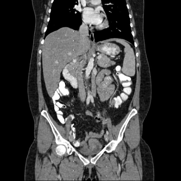
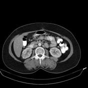
Duplicated left sided IVC is seen as a continuation of left common iliac vein, crossing anterior to aorta at the level of renal vein to join the right sided vena cava.
Case Discussion
The findings are consistent with duplication of inferior vena cava, a relatively rare vascular anomaly.


 Unable to process the form. Check for errors and try again.