
Presentation
This patient presented to the ED with nonspecific abdominal pain.
Patient Data
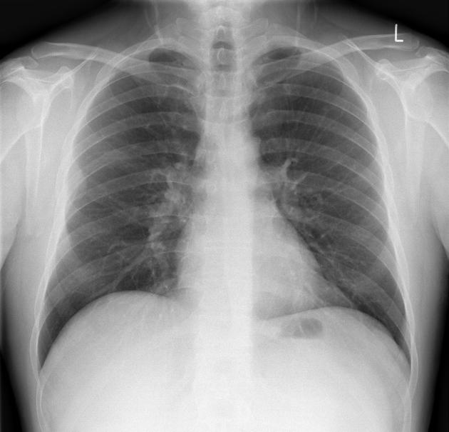
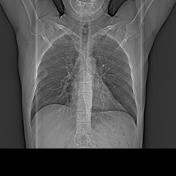
Erect CXR to look for free gas under the diaphragm
No free gas under the diaphragm. In the right midzone, there is a sharply circumscribed peripheral lesion forming obtuse angles with the chest wall. Lungs are clear. Visible bones appear normal.
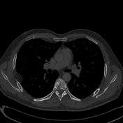

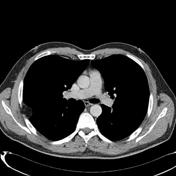
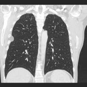
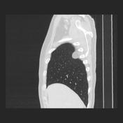

Performed to investigate the lesion seen on chest radiograph (considered incidental).
There is a non-enhancing fat attenuation pleural based lesion in the right posterolateral hemithorax. The lungs are otherwise normal in appearance and there is no pneumoperitoneum.
Findings are in keeping with a pleural lipoma.
Case Discussion
As is usually the case, this pleural lipoma was incidentally found on chest radiograph.
No follow up was recommended.


 Unable to process the form. Check for errors and try again.