
Presentation
Micro-opthalmia and leukocoria.
Patient Data
Age: 3 months
Gender: Male
From the case:
Persistent hyperplastic primary vitreous

Download
Info
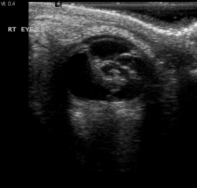
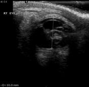
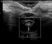
The right globe is small and contains a complex lesion with a vascular stalk that connects it to the posterior internal surface of the globe.
Case Discussion
The right orbit is small (microphthalmia) and contains a complex central lesion in the anterior portion of the vitreous. It is connected to the midline posterior globe by a vascular stalk.
Features are of persistent hyperplastic primary vitreous.


 Unable to process the form. Check for errors and try again.