
Presentation
High output cardiac failure. Failed attempt at medical control and weaning from ventilator.
Patient Data


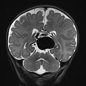
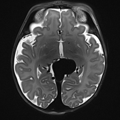
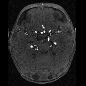
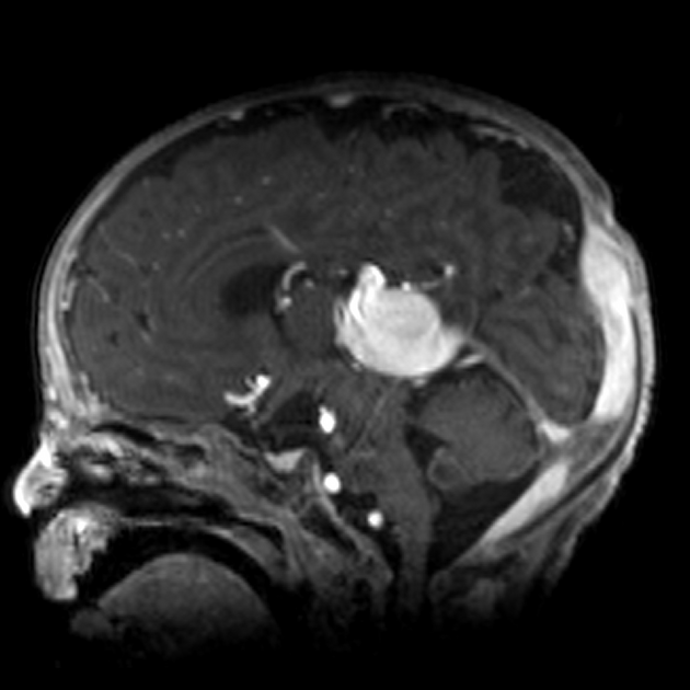
A large vascular structure is noted in the midline below the splenium of the corpus callosum, draining into the transverse sinuses. The anterior cerebral arteries are very large as are the posterior cerebral arteries. Features are characteristic of a vein of Galen malformation.

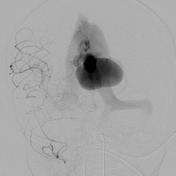

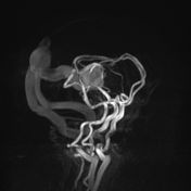
The vein of Galen malformation fills via the posterior communicating artery (PCOM) and right posterior cerebral artery (PCA) branches and by right and left anterior cerebral artery (ACA) subfornical branches forming a complete limbic arch.
The distal right ACA demonstrates a large high-flow fistula into the vein of Galen malformation (VGAM).
Note that the contrast bolus shunts through the malformation so quickly that it reappears as a bolus a few seconds later after a complete cardiac cycle.
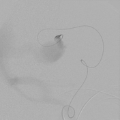
Attempts to coil the artery at the fistula to allow subsequent glue failed, due to the coil prolapsing into VGAM. Hence the malformation itself was embolized with platinum coils to provide a basket to allow subsequent occlusion at fistula.
Distal right ACA then 1:1 NBCA - with good occlusion and penetration to the fistulas, but no penetration beyond coil mass in the malformation.
Distal right PCA choroidal branch also embolized (not shown) 1:1.8 NBCA - after coil embolization from malformation into terminal artery. PCA cortical branches arise within mm's of this large branch - despite slow injection, reflux into a small adjacent branch.
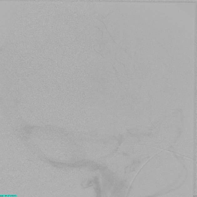
At the end of the procedure, there was still flow in the malformation but this was decreased, and an increase in blood pressure was noted during treatment consistent with improved cardiac function.
Angiography courtesy of Prof. Peter Mitchell.
Case Discussion
Typical appearances of a vein of Galen malformation.
Unfortunately no further followup is available.


 Unable to process the form. Check for errors and try again.