
Presentation
Abdominal pain and constipation. Recurrent abdominal cystic lesion with history of rupture and previous operations.
Patient Data
Age: 70 years
Gender: Female
From the case:
Calcified cystic mesenteric lymphangioma


Download
Info

- A large right-sided multi-locular cystic mesenteric lesion is seen filling the right lumbar and iliac regions. The lesion shows rather dense contents with thin internal septations. The lesion shows dense coarse calcifications of its wall mainly posteriorly. No mural solid component is noted. the lesion shows no evident post-contrast enhancement. The lesion measures 14 X 11 X 7.5 cm in its main CC and axial diameters respectively.
- No enlarged abdominal or pelvic lymph nodes.
- Normal liver and spleen with no definite focal lesions.
- The retro-peritoneal structures including the pancreas and both kidneys are within normal.
- Infra-umbilical hernia with small bowel loops and mesenteric fat is noted.
From the case:
Calcified cystic mesenteric lymphangioma
Download
Info
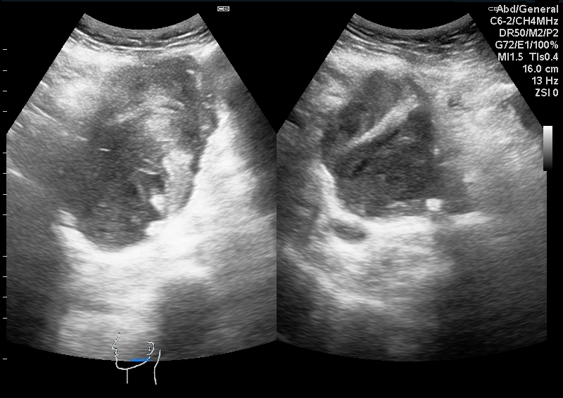
US shows multilocular cystic lesion with septations and thick tenaceous contents demonstrating enhanced through transmission. Wall calcifications with posterior shadowing is noted as well.


 Unable to process the form. Check for errors and try again.