
Presentation
No history provided.
Patient Data
Age: Adult
From the case:
Cerebral arteriovenous malformation


Download
Info
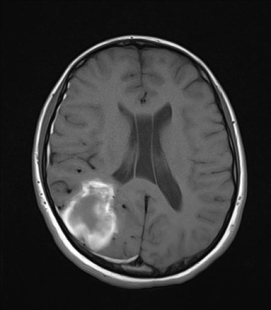
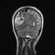
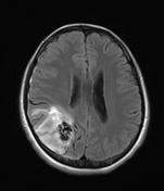
A hemorrhagic mass is seen in the T1W images. Typical "salt and pepper" appearance due to multiple flow voids is seen in the T2W series. This is diagnostic for brain arteriovenous malformation (AVM).
Note the presence of a draining vein coursing to the superior sagittal sinus. There is also an aneurysm near the nidus.
Also note the small right sided subdural hematoma.
Case Discussion
Cerebral arteriovenous malformations (AVMs) are the most common symptomatic cerebrovascular malformations. Their most common presentation is intraparenchymal hematoma.
Large AVMs are typically wedge-shaped lesions located in the watershed area, with their apex toward the ventricle.


 Unable to process the form. Check for errors and try again.