
Presentation
Abdominal pain and discomfort
Patient Data
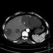


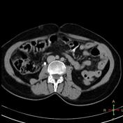

Multiple hepatic focal lesions the one involving the left lobe is giant and they are displaying typical incomplete closed iris sign due to centripetal contrast filling.

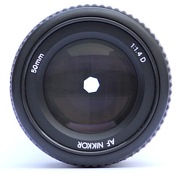

Open and closed camera aperture demonstrating the iris sign.
Author: KoeppiK
Source URL: https://commons.wikimedia.org/wiki/File:Lenses_with_different_apertures.jpg
Modifications: Square crop, remove text.
Licence: This file is licensed under the Creative Commons Attribution-Share Alike 4.0 International, 3.0 Unported, 2.5 Generic, 2.0 Generic and 1.0 Generic license.
If you believe your copyright has been infringed, please write to license@radiopaedia.org, explaining why you believe this is so.
Case Discussion
Giant hepatic hemangiomas are associated with Kasabach-Merritt syndrome. Hemangiomas are multiple in 10% of cases.


 Unable to process the form. Check for errors and try again.