
Presentation
Fever, dyspnea, left side chest pain and productive cough.
Patient Data
Age: 75 years
Gender: Male
From the case:
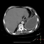
Thoracic empyema


Download
Info
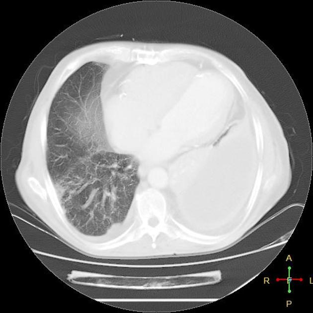
Large left pleural effusion that shows thick enhancing pleural lining with typical split pleura sign. Compression upon the left lung and mediastinal lymphadenopathies are seen.
Case Discussion
Split pleura sign can differentiate between empyema and lung abscess.
Unlike virtually all other chest imaging, with a clinical concern regarding the pleura, particularly in clinical suspicion of empyema, performing the study at 70 seconds, tends to give the best diagnostic images.


 Unable to process the form. Check for errors and try again.