
Presentation
Epileptic fits.
Patient Data
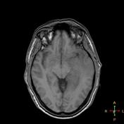

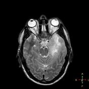
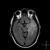
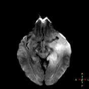
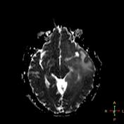
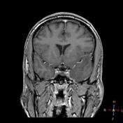
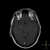

Left temporal subcortical ill defined lesion that blurs the grey/white matter junction of the medial temporal lobe and doesn't cause any mass effect or any cortical destruction. It displays slightly low signal in T1 and bright signal in T2 and FLAIR without significant enhancement in post contrast study. The lesion exhibits bright signal in DWI and ADC suggesting T2 shine through effect and excludes recent infarction.
Diagnosis: Gliomatosis cerebri
Case Discussion
The differential includes viral (herpes) encephalitis, progressive multifocal leukoencephalopathy (PML), lymphoma, demyelination and infarction. The patient underwent craniotomy and biopsy by which the diagnosis of gliomatosis cerebri WHO grade III was made.
Importantly, whereas gliomatosis was previously considered a distinct entity, since the 2016 update to the WHO classification of CNS tumors it is now merely thought of as a growth pattern.


 Unable to process the form. Check for errors and try again.