
Presentation
IV drug user. Now complaining of fever, productive cough and chest pain.
Patient Data
Age: 35 years
Gender: Male


Download
Info
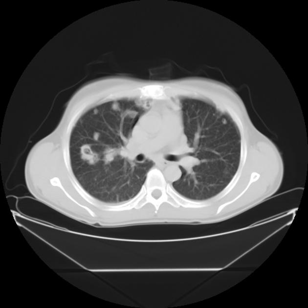
Both lungs are studded with numerous variable sized cavitating nodules that appear to colese in both lower lobes.
Bilateral minimal pleural effusion.
From the case:
Septic pulmonary emboli
Download
Info
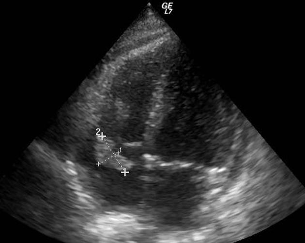
A mass is seen attached to lateral leaflet of tricuspid valve measuring about 13 mm X 25 mm on echocardiography.
Case Discussion
Bilateral pulmonary cavitating nodules. In light of the patient’s history and echocardiographic data, these findings represent pulmonary septic emboli as a complication of IV drug use associated infective endocarditis (infected tricuspid vegetation).


 Unable to process the form. Check for errors and try again.