
Presentation
Ongoing right L4 radiculopathy. Otherwise well.
Patient Data
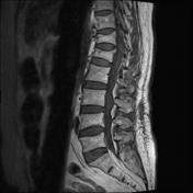


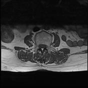
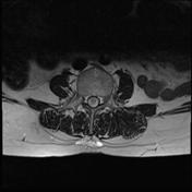

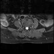

Relatively subtle T1 isointense, T2 hyperintense lesion at the L3 vertebral body level in the central canal, which enhances avidly following contrast.
The relatively subtle findings on non-contrast sequences become obvious following contrast. The nature and relationship to the cauda equina and nerve roots are very well delineated on the post-gad sequences.
Case Discussion
Incidental finding of a intrathecal, extramedullary mass. The differential includes; schwannoma, neurofibroma or a metastasis. The former is in the most likely in this case, given the imaging characteristics. Note the similarity in appearances, especially on the post contrast sequences to a schwannoma at the IAM - avidly enhancing with small cystic areas of degeneration within.
The appearances were static on a repeat scan 5 months later.


 Unable to process the form. Check for errors and try again.