
Presentation
Trauma.
Patient Data
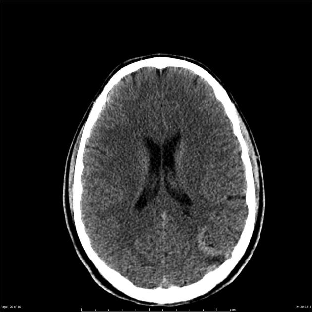
Noncontrast axial images through the brain have been obtained demonstrating a serpiginous hyperdensity is in the left parietal region.
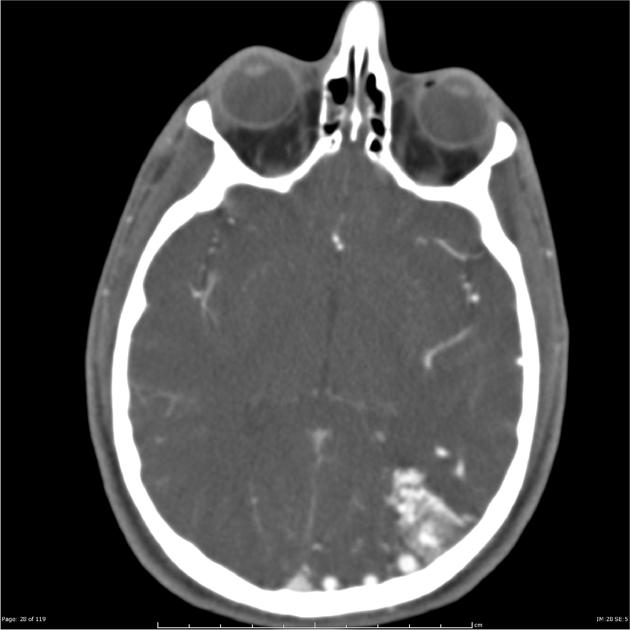
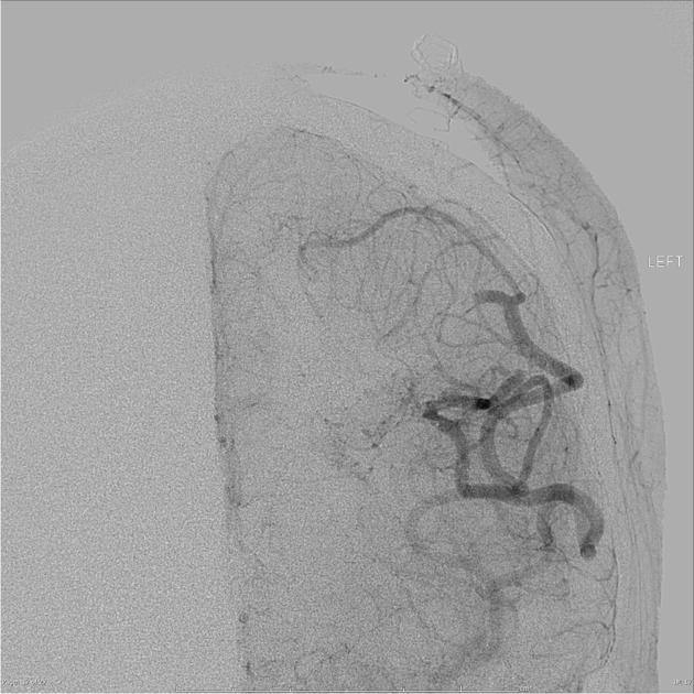
A relatively compact arteriovenous malformation is located superficially in the parieto-occipital region on the left, measuring 31 x 18 x 32 mm. It receives most of its supply from the left middle cerebral artery branches, with further contribution from the left posterior cerebral artery. No convincing external carotid supply can be demonstrated. Venous drainage appears predominantly (exclusively?) superficial, with a very large vein coursing posteriorly to drain into the superior sagittal sinus. There is no evidence of hemorrhage or thrombosis or aneurysm formation.
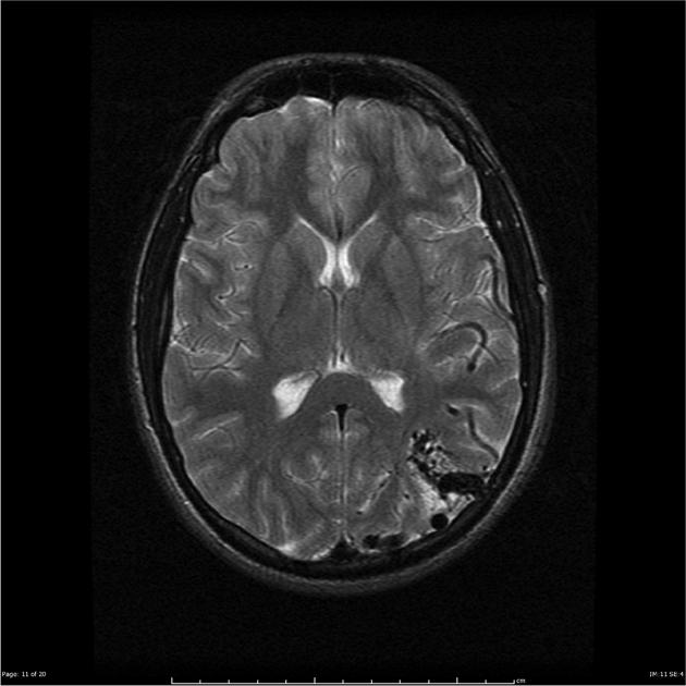
Note the presence of flow voids in the AVM.


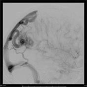
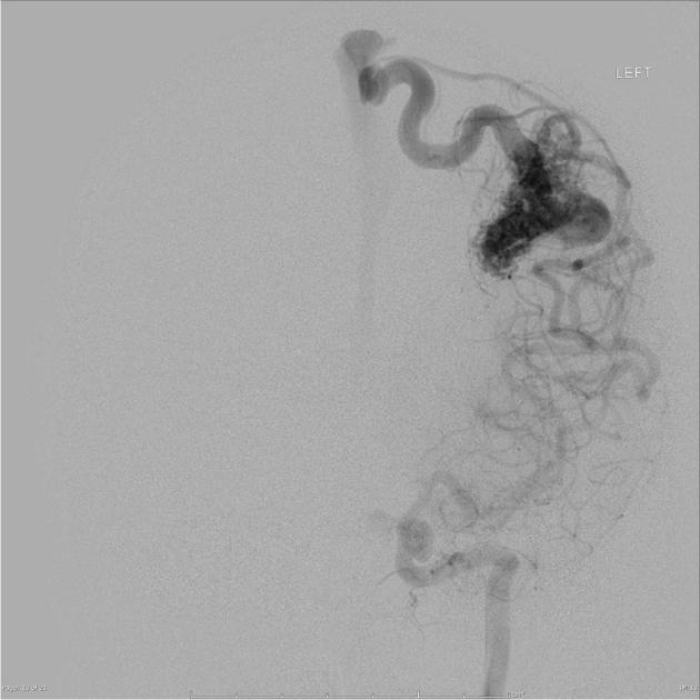
An arteriovenous malformation at the junction of left parietal and occipital lobes as a nidus measuring just over 3 cm diameter. Principal blood supply is from enlarged parietal and posterior temporal branches of the left MCA with some contribution from left PCA branches. No right sided supply and no external carotid supply. Venous drainage is via a large vein draining to the superior sagittal sinus and a smaller superficial vein draining to the left sigmoid sinus. No arterial flow related aneurysms.
Conclusion:Left parieto occipital AVM ( Spetzler Martin Grade 3).
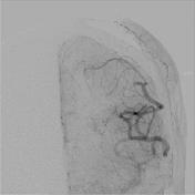

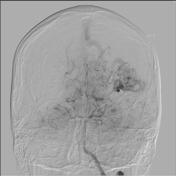
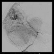
Large defect in parenchymal phase marks site of resection of the AVM - with no definite AV shunting. Large angular and posterior temporal branches of left MCA - sluggish flow, up to local occlusion of the branches at the level shown previously to contain the AVM. The only area of suspicion for residual nidus was at the anterior margin on the lateral view - with some abnormal vessels, but without definite shunting. No ECA or contralateral supply; the VA-PCA supply has also been removed.
Case Discussion
The patient underwent a satisfactory post-operative resection.


 Unable to process the form. Check for errors and try again.