
Presentation
Epigastric fullness.
Patient Data
Age: 25 years
Gender: Female
From the case:
Pulmonary hydatid cyst

Download
Info

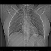
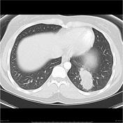
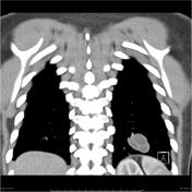
There is a lobulated mass lesion in the left lower lobe, abutting the left hemidiaphragm. The lesion measures 26 x 45 x 33mm. Internally, the lesion is predominantly fluid density, and there are internal septations. No cavitation or wall calcification.
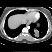
Note made of a partially visualized large cystic lesion within the left lobe of the liver, which measures 6.7 x 10.0cm.
The liver and left lower lobe lung lesion are most likely hydatid cysts.
Case Discussion
Left lower lobe lobectomy confirmed a hydatid cyst.


 Unable to process the form. Check for errors and try again.