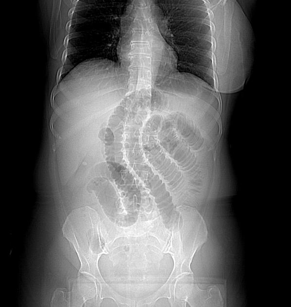
Incarcerated Spigelian hernia with mechanical small bowel obstruction
Diagnosis certain
Presentation
50-year-old woman with right modified radical mastectomy and chemo-radiotherapy 6 years previously is now presenting with severe abdominal pain, distension and vomiting.
Patient Data
Age: 50 years
Gender: Female
Download
Info

Frontal radiograph shows dilated small bowel loops with an abrupt cut-off at the right iliac region. No radiographic signs of pneumoperitoneum.
A dense calcific shadowing is seen in the right upper quadrant region representing a gallstone.
{"current_user":null,"step_through_annotations":true,"access":{"can_edit":false,"can_download":true,"can_toggle_annotations":true,"can_feature":false,"can_examine_pipeline_reports":false,"can_pin":false},"extraPropsURL":"/studies/26783/annotated_viewer_json?c=1669266558\u0026lang=us"}
- Marked dilatation of the small bowel loops reaching up to 4 cm in its maximal diameter with an abrupt transition at the distal ileum within the right iliac region. The distal ileal loops are seen entangled within the intermuscular plane of the lower right anterior abdominal wall being between the external oblique and transverse abdominis muscles passing through a defect in the internal oblique aponeurosis measuring 2.1 X 1.6 cm. The hernial sac lies just beneath the intact aponeurosis of the external oblique muscle. The most distal ileal loops appear collapsed.
- Mild peri-hepatic and pelvic ascites. The liver is mildly enlarged with smooth regular outline, no definite focal hepatic lesion or intrahepatic biliary dilatation.
- The gallbladder shows a 1 cm gallstone.
Case Discussion
Incarcerated Spigelian hernia with small bowel obstruction.


 Unable to process the form. Check for errors and try again.