
Presentation
3-month history of urinary difficulties culminating in acute admission with urinary and fecal incontinence.
Patient Data


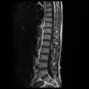
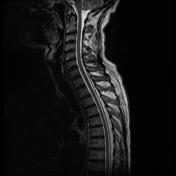
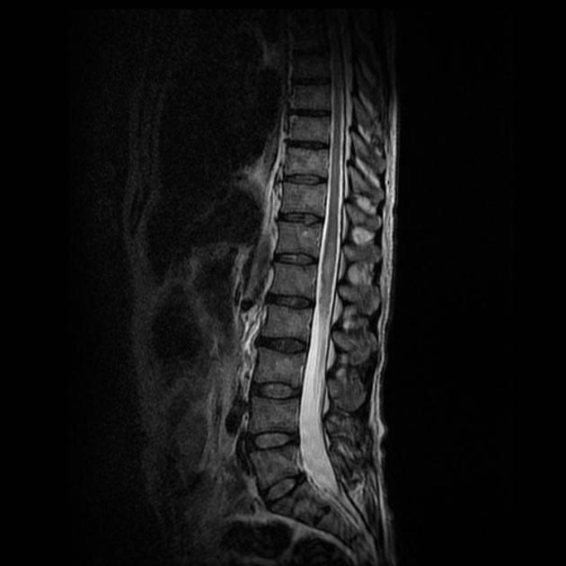
T2 images show a hyperintense lesion in the conus, with slight expansion. This enhances following contrast administration. The intense solid-like enhancement does not favor demyelination or other inflammatory disorder. A tumor such as ependymoma was considered at the time of imaging.
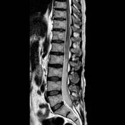
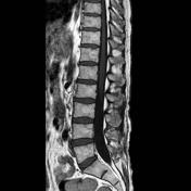
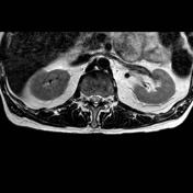

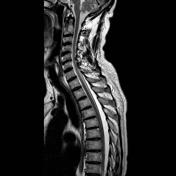

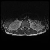
On this follow up scan, the conus is even more hyperintense and expanded. The area of enhancement has also increased, and note the increased vascularity of the pial vessels leading up to the lesion. On the axials, a small non-enchancing necrotic area has developed on the right side of the lesion. New enhancement of the cauda roots is also seen, suggesting leptomeningeal dissemination.
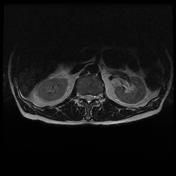
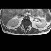
An interesting associated finding is thoracic epidural lipomatosis secondary to the high dose steroids. The proliferation of epidural fat dorsal to the thoracic cord causes the CSF anterior to the cord to be effaced (compare with original study).
Case Discussion
The appearances following the second scan were strongly in favor of an intrinsic cord tumor (CT body was negative). The patient went for a debulking, and the histology showed glioblastoma (GBM). This is an example of a common tumor in an uncommon location. In retrospect, the imaging findings were all those expected for a GBM if seen in the brain.


 Unable to process the form. Check for errors and try again.