
Presentation
Abdominal pain - 2 days
Patient Data
Age: 19 years
Gender: Male
From the case:
Appendicitis - perforated


Download
Info

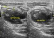
Appendix is visualized in its entire length. There is a focal area of loss of gut signature sign in the appendix consistent with perforation and an adjacent collection. On compression, echoes appear to move from appendix perforation to adjacent collection.
Echogenic, inflammed periappendiceal fat and reactive nodes are present.
Appendiceal wall vascularity is preserved.
No fecolith is noted.
Case Discussion
Findings favor appendicitis with perforation and local collection.


 Unable to process the form. Check for errors and try again.