
Presentation
Headache.
Patient Data


Hyperdense lesion on non contrast scan. See MR report.

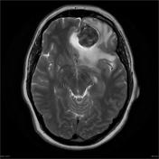


T2 hypointense T1 isointense vividly enhancing mass applied to the floor of the anterior cranial fossa left of midline has dimensions of 3.4 x 3.6 x 4.6 cm. There is extensive vasogenic edema through the left frontal lobe and into the genu of the corpus callosum. The anterior aspect of the falx is deviated toward the right, with 12 mm subfalcine herniation. No uncal herniation. No evidence of extension of tumor into orbit, ethmoid or nasal cavity. No further intracranial mass or site of pathological contrast enhancement. A few tiny T2 hyperintense white matter foci are compatible with patient age. No hydrocephalus.


Case Discussion
The sections show a moderately cellular tumor. The tumor forms fascicles and bundles, intermixed with collagenous tissue in the background. No whorls are seen. The tumor cells have ovoid nuclei with no nuclear pleomorphism. Occasional mitoses are noted (less than 4 per 10 high power fields). Scattered staghorn type blood vessels are present. There is one area of brain invasion, with tumor infiltrating into the neuropil.
DIAGNOSIS: Brain tumor: Solitary fibrous tumor.
Comment: No grading has been given in the WHO classification. Most behave in a benign fashion. However, there is brain invasion in this biopsy and its significance is unclear. No other adverse histological features are otherwise seen.


 Unable to process the form. Check for errors and try again.