
Presentation
Dysphagia, vomiting, chest pain and weight loss.
Patient Data
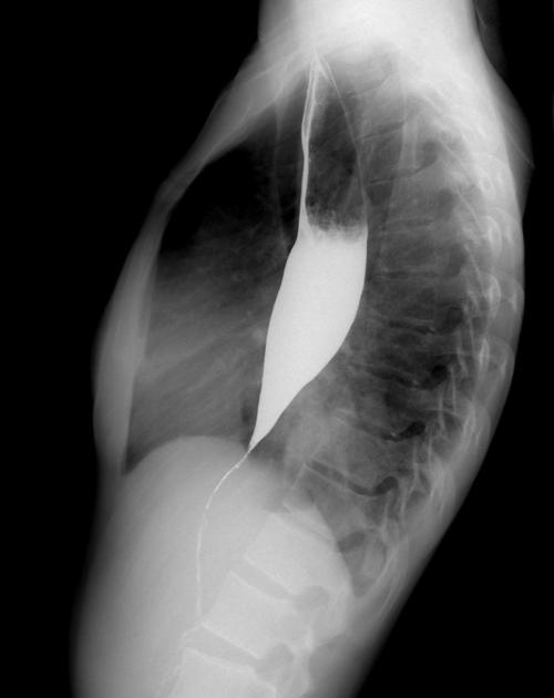
The upper and mid thoracic esophageous is dilated with absent 1ry and 2ry peristaltic waves all through the study as well as few tertiary contractions and peaked tapered distal esophageous and GEJ that failed to dilate in response to swallowing.
Normal mucosal pattern with no irregularities or ulceration.
No extrinsic compressions.
Free flow of the Barium bolus along the pharynx & cervical esophagus with no evidence of obstruction, strictures or filling defects.
No evidence of reflux or hiatus hernia.
Right lower lung lobe medial segment paracardiac wedge shaped patch of opacity with air bronchogram within.
Case Discussion
The above described findings are those of esophageal achalasia with right lower lung lobe medial segment aspiration pneumonia.


 Unable to process the form. Check for errors and try again.