
Patient Data
Age: 45 years
Gender: Male
Download
Info
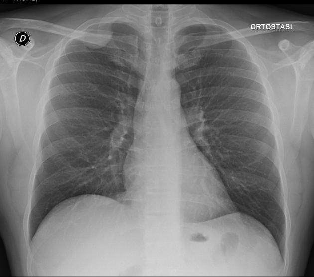
Chest radiography showed a homogeneous subpleural rounded shadow at the right upper lobe.


Download
Info
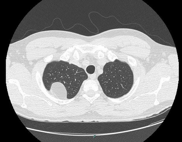
CT revealed a well-circumscribed lesion with mean attenuation value -115 HU, in contact with the posterior third rib arc and at an obtuse angle with the chest wall.
Case Discussion
Pleural lipomas originate from the submesothelial parietal pleura and extend into the subpleural, pleura, or extrapleural space. Most patients are asymptomatic and the tumor is usually discovered incidentally on chest radiography. CT allows a definitive diagnosis when the mass demonstrates a homogeneous fat attenuation (-50 to -150 HU).


 Unable to process the form. Check for errors and try again.